- 1. Introduction of DTCT in Idar-Oberstein
- 2. What is Digital Tomosynthesis Computed Tomography (DTCT)?
- 3. Applications of DTCT in veterinary medicine
- 4. Advantages of DTCT over traditional imaging techniques
- 5. The process of DTCT imaging in animal practice
- 6. Case studies: DTCT in practice
- 7. Expert Opinions: The Impact of DTCT in Veterinary Medicine
- Advantages of DTCT
- Areas of application of DTCT
- 8. Challenges and considerations in implementing the DTCT
- 9. Future trends in veterinary imaging
- 10. Practical tips for veterinarians when using the DTCT
- Frequently asked questions about our DTCT in Idar-Oberstein
- 11. Conclusion
1. Introduction of DTCT in Idar-Oberstein
Digital tomosynthesis computed tomography (DTCT), also known as digital volume tomography (DVT), represents a significant advance in medical imaging, particularly in the fields of dentistry and veterinary medicine. This technology enables the creation of high-resolution, three-dimensional images through a sophisticated combination of an X-ray tube and a flat-panel detector that rotate around the patient. Compared to traditional CT scans, DTCT provides an unmatched level of detail, making it an indispensable tool in the diagnosis and treatment of a variety of animal diseases.
In this article, we will explore the various aspects of DTCT, its applications in veterinary medicine, and the advantages it offers over traditional imaging techniques. We also discuss expert opinions, case studies and future trends in this area.

2. What is Digital Tomosynthesis Computed Tomography (DTCT)?
What is DTCT in Idar-Oberstein?
DTCT is a state-of-the-art imaging technique that combines elements of digital tomosynthesis and computed tomography to create detailed three-dimensional images. It is particularly useful in areas where precise imaging is critical, such as dental and oral surgery.
How is DTCT different from traditional CT scans?
While both DTCT and traditional CT scans provide three-dimensional images, DTCT offers significantly higher resolution, making it possible to detect minute details that might be missed CT imaging Additionally, DTCT typically uses a lower dose of radiation, making it a safer option, especially for repeated applications.
Technical aspects of the DTCT in Idar-Oberstein: How does it work?
The DTCT works by rotating a combination of an X-ray tube and flat detector around the patient. As the X-ray tube emits radiation, the detector captures multiple images from different angles, which are then reconstructed into a detailed 3D model. This model can be examined from different perspectives, providing a comprehensive view of the area of interest.
3. Applications of DTCT in veterinary medicine
DTCT in dental imaging in animals
One of the main applications of DTCT in veterinary medicine is dental imaging. In animals such as dogs and cats, accurate dental imaging is critical to diagnose conditions such as tooth decay, periodontal disease and oral tumors. The DTCT allows veterinarians to visualize the teeth, roots and surrounding bone tissue in exceptional detail.
Use in oral and maxillofacial surgery
In cases where oral or maxillofacial surgery is required, DTCT provides surgeons with detailed preoperative images that help them plan and perform procedures more precisely. This is particularly beneficial for complex surgeries involving the jaw or facial bones.
Applications for ENT diseases
The DTCT is also extremely effective in diagnosing and treating ENT diseases in animals. For example, it can be used to assess ear infections, nasal congestion, or other head and neck conditions, providing the details necessary for accurate treatment.
Special cases: imaging for small animals such as rodents and rabbits
Small animals such as rodents and rabbits present particular challenges in veterinary imaging because they are very small. The DTCT is ideal for these cases because it provides detailed images that allow veterinarians to diagnose conditions that might otherwise be difficult to detect.
4. Advantages of DTCT over traditional imaging techniques
Higher resolution and detail
One of the greatest advantages of DTCT is its ability to produce images with much higher resolution than traditional imaging techniques. This higher level of detail is particularly important in dental imaging, where small anatomical structures must be clearly displayed.
Reduced radiation exposure
DTCT typically uses a lower dose of radiation than traditional CT scans. This makes it a safer option for animals, especially those that require multiple imaging sessions.
Increased diagnostic accuracy
The high resolution and three-dimensional nature of DTCT images enable more accurate diagnoses, leading to better treatment outcomes. Veterinarians can detect and treat diseases earlier and more effectively.
5. The process of DTCT imaging in animal practice
Preparing the animal for DTCT
Before a DTCT examination, the animal may need to be sedated or anesthetized to ensure that it remains calm during the procedure. This helps avoid motion artifacts and ensures images are of the highest quality.
The imaging process: what happens step by step
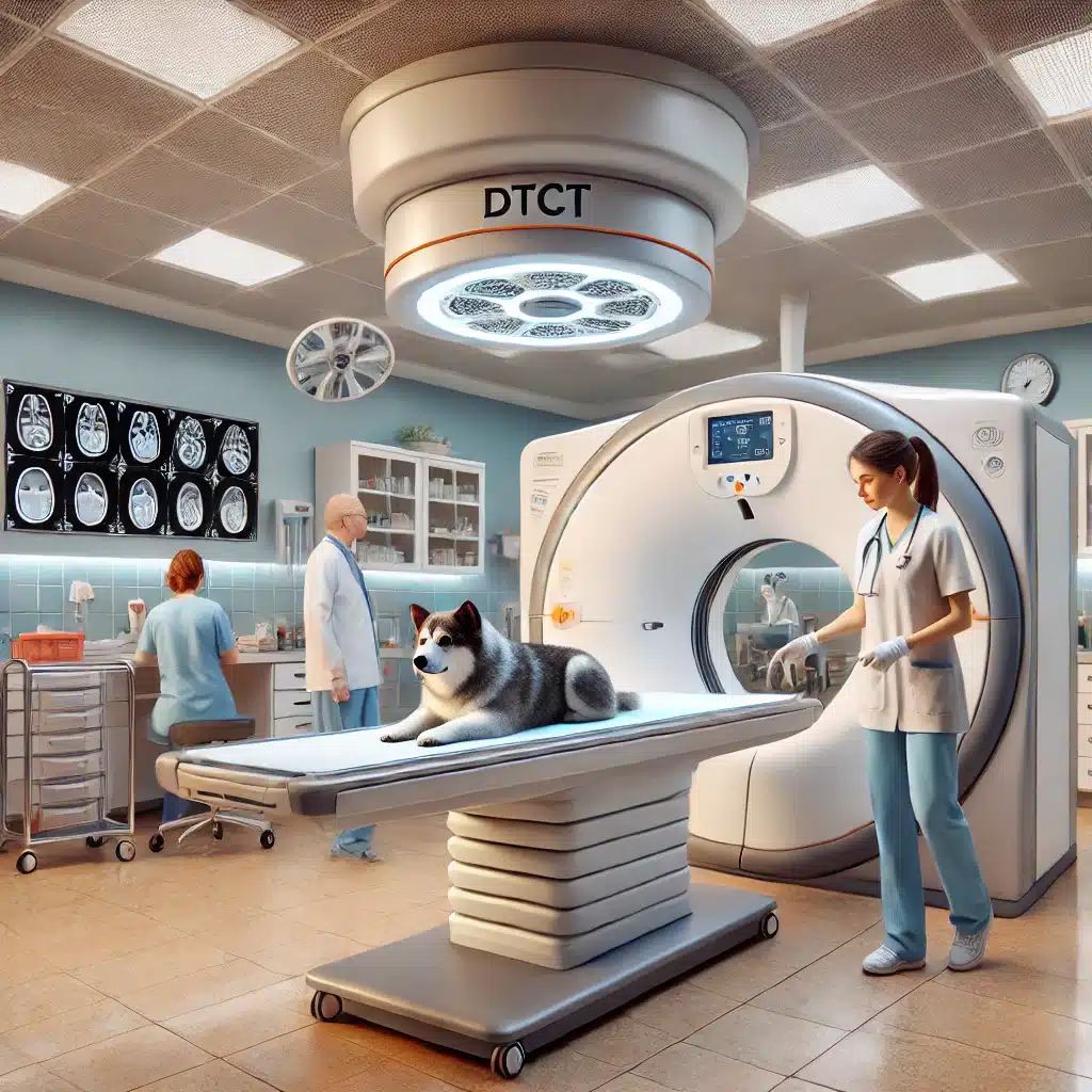
During the DTCT procedure, the animal is positioned on the imaging table and the X-ray tube and detector rotate around the animal to capture multiple images. These images are then processed and reconstructed into a 3D model that the veterinarian can analyze.
Post-imaging analysis and interpretation
Once the DTCT images are available, they will be reviewed by a veterinarian or specialist. The detailed 3D images allow for a thorough examination of the area of interest, and the results are used to make treatment decisions.
6. Case studies: DTCT in practice
Case study 1: Dental imaging in a dog
A 7-year-old Labrador Retriever showed signs of mouth pain. Conventional radiographs were unremarkable, so a DTCT scan was performed. The scan revealed a tooth root fracture that was not visible on the standard images, resulting in a successful extraction and the dog being pain free.
Case study 2: Jaw surgery on a cat
A Persian cat needed surgery for a broken jaw. The DTCT provided the surgical team with detailed images of the fracture, allowing for precise surgical planning and a successful outcome.
Case study 3: ENT diagnostics in a rabbit
A rabbit with chronic nasal discharge underwent DTCT imaging, which revealed a nasal tumor. The detailed images allowed the veterinary team to plan and carry out a successful surgical removal.
7. Expert Opinions: The Impact of DTCT in Veterinary Medicine
Veterinary experts have noted that DTCT has revolutionized diagnostic imaging in animals. Dr. Jane Smith, a veterinary radiologist, says: “DTCT gives us the opportunity to see things that were invisible with traditional imaging techniques. It has significantly improved our diagnostic capabilities and treatment outcomes.”
DTCT in Idar-Oberstein: Advantages and areas of application
Advantages of DTCT
- High resolution: Enables extremely detailed 3D images that make even the smallest structures visible.
- Low radiation exposure: Gentle examination thanks to reduced radiation dose compared to conventional CT scans.
- Precise diagnoses: Improved diagnostics through accurate representation of teeth, jaws and other structures.
- Versatile uses: Ideal for various applications such as dentistry, oral surgery and otolaryngology.
Areas of application of DTCT
- Dentistry: Diagnosis of dental diseases, such as tooth decay and root inflammation, with high precision.
- Oral and maxillofacial surgery: Support in the planning and implementation of complex surgical procedures in the jaw area.
- Otolaryngology: Detailed imaging for ear infections, nose problems and tumors in the head and neck area.
- Small pets: Particularly suitable for examining small animals such as rodents and rabbits, where precision is particularly important.
8. Challenges and considerations in implementing the DTCT
Cost considerations and accessibility
Although DTCT offers many benefits, the cost of the equipment and the need for specialized training can be a hurdle for some veterinary practices. However, these costs are expected to fall as the technology becomes more widespread.
Training and expertise for accurate interpretation
Interpretation of DTCT images requires specialized training and experience. Veterinarians must be trained to read these detailed images correctly to avoid misdiagnosis and ensure the best possible care for patients.
Ethical considerations and animal safety
As with any medical procedure, the safety and well-being of the animal is paramount. Because DTCT involves radiation exposure, it is important to weigh the benefits of the exam against the potential risks.
9. Future trends in veterinary imaging
New technologies and their potential impact
Advances in imaging technology continue to evolve and new methods are being developed that could further improve diagnostic capabilities in veterinary medicine. Innovations such as AI-supported image analysis are already in the starting blocks.
The future of DTCT in veterinary practice
As DTCT technology becomes more accessible, it is expected to become a standard tool in veterinary imaging. Further research and development will further refine its applications and make it an even more powerful diagnostic tool.
10. Practical tips for veterinarians when using the DTCT
Best practices for optimal image results
To achieve the best results with DTCT, it is important to follow best practices, including proper positioning of the patient, minimizing movement during the scan, and using the appropriate imaging settings for each case.
Common pitfalls and how to avoid them
A common pitfall with DTCT imaging is the misinterpretation of artifacts as pathology. Veterinarians should be trained to distinguish between true anatomical structures and artifacts.
Frequently asked questions about our DTCT in Idar-Oberstein
What is a digital tomosynthesis computed tomography (DTCT) and how does it work?
A digital tomosynthesis computed tomography (DTCT) is a state-of-the-art imaging device that creates high-resolution, three-dimensional images. In contrast to conventional CT scanners, the DTCT uses a combination of a rotating X-ray tube and a flat detector. These rotate around the patient and take images from different angles. These images are then digitally assembled into a detailed 3D model of the area examined. DTCT allows us to precisely image even the smallest structures in the body, such as teeth or bones. This accuracy is particularly important in dentistry, oral surgery and otolaryngology, as it enables early and accurate diagnosis as well as optimal treatment planning.
What advantages does DTCT offer compared to conventional X-rays?
The DTCT offers numerous advantages over conventional X-rays. Firstly, the image quality is significantly higher because the DTCT delivers three-dimensional images with a much higher resolution. This allows us to see even the smallest anatomical details that may not be visible on traditional x-rays. Secondly, the radiation exposure from a DTCT is usually lower than from a conventional CT scan, which is particularly important when repeat examinations are required. Thirdly, 3D imaging allows for more precise diagnosis and better planning of interventions, as we can view the affected area from different angles. Overall, DTCT offers a safer, more detailed and gentler imaging option.
For which animals is the DTCT suitable and what areas of application are there?
Our DTCT in Idar-Oberstein is suitable for a wide range of animals, including dogs, cats, rabbits and even small rodents. The DTCT has a wide range of applications in veterinary medicine. A main area of application is dentistry, where DTCT helps to diagnose dental diseases such as tooth decay, tooth root inflammation or tumors in the oral cavity. In addition, DTCT is used in maxillofacial surgery to detect complex fractures or other structural problems and to plan precise procedures. DTCT is also of great use in otolaryngology, for example for diagnosing ear infections, nose problems or tumors in the head and neck area. Thanks to the high image quality and the ability to accurately display even the smallest structures, DTCT is suitable for almost all diagnostic and therapeutic questions.
How does a DTCT examination work for my animal and what do I have to consider?
The process of a DTCT examination is usually uncomplicated and well tolerated by your animal. Before the scan, the animal may be lightly sedated or placed under anesthesia to ensure that it remains still during the scan. This is important to avoid motion artifacts and obtain high-quality images. The animal is then placed on the examination table and the DTCT scanner begins rotating around the animal, taking images from different angles. This process only takes a few minutes. After the examination, your animal can usually go home quickly as soon as it has completely woken up from the sedation or anesthesia. It is important that you do not give your pet any food before the examination if sedation is planned, and after the examination your animal should rest in a quiet environment.
Is the DTCT examination safe for my animal and what are the risks?
Yes, the DTCT examination is safe for your animal. The DTCT uses significantly lower levels of radiation than traditional CT scanners, making it particularly safe for animals that may require repeat examinations. The sedation or anesthesia sometimes required for the exam will be carefully monitored to ensure your pet tolerates the procedure well. As with any medical examination, there are small risks, but these are usually minimal and are greatly outweighed by the benefits of accurate diagnosis and resulting better treatment. Our experienced team will ensure that all necessary precautions are taken to ensure your animal's safety and well-being throughout the examination.
11. Conclusion
Digital tomosynthesis computed tomography (DTCT) has become an essential tool in veterinary practice, providing unprecedented image detail that improves diagnostic accuracy and treatment outcomes. As technology advances, DTCT is expected to play an even more central role in veterinary medicine, helping to further improve the care and quality of life of animals.
In our practice in Idar-Oberstein we use the advanced digital tomosynthesis computer tomography (DTCT), also known as digital volume tomography (DVT). With our DTCT in Idar-Oberstein, we offer precise and detailed imaging that allows us to display even the smallest structures such as teeth and jawbones with the highest level of accuracy. The DTCT in Idar-Oberstein is ideally suited for various veterinary applications, be it in dentistry, complex surgical procedures or ENT medicine.
Our DTCT in Idar-Oberstein enables significantly improved diagnostics compared to conventional imaging methods, as it delivers images with higher resolution and lower radiation exposure. This allows us in Idar-Oberstein to make an exact and reliable diagnosis, which forms the basis for optimal treatment for your animals. Regardless of whether it is a dog, cat, rabbit or rodent – the DTCT in Idar-Oberstein is versatile and offers the right solution for every animal.
Thanks to the DTCT in Idar-Oberstein, we can now gain deeper insights in cases where a simple X-ray image is not enough, thus enabling more precise treatment planning. Our DTCT in Idar-Oberstein is an integral part of our practice and stands for state-of-the-art imaging technology that focuses on the well-being of your animal.
Trust our DTCT in Idar-Oberstein when it comes to your animal’s health. With the DTCT in Idar-Oberstein, we offer you and your animal a state-of-the-art, safe and precise diagnostic option that meets the highest standards.
By using the DTCT in Idar-Oberstein, we are able to plan and carry out treatments and procedures even more precisely. The detailed, three-dimensional images generated by our DTCT in Idar-Oberstein allow us to view complex anatomical structures from different perspectives. This is particularly important in dentistry, where the smallest changes or illnesses can be detected and treated early.
Our DTCT in Idar-Oberstein also offers significant advantages in ENT medicine. Whether diagnosing ear infections, nose problems or other diseases in the head and neck area - the DTCT in Idar-Oberstein provides the necessary precise information to ensure the best possible treatment. The high resolution of the DTCT in Idar-Oberstein allows us to capture even the smallest details that are often overlooked on conventional X-ray images.
Another advantage of DTCT in Idar-Oberstein is the low radiation exposure, which is particularly important for repeated examinations. This makes the DTCT in Idar-Oberstein a safe choice for all animal species and age groups. Our patients benefit from the modern technology of the DTCT in Idar-Oberstein, which enables gentle and at the same time extremely effective diagnostics.
If you are looking for a reliable and advanced imaging method in Idar-Oberstein, then our DTCT is the optimal solution. The DTCT in Idar-Oberstein is not only a technological advance, but also a decisive step towards even better animal health. Trust in the precise diagnostics that our DTCT in Idar-Oberstein offers and ensure the best care for your animal.
With the DTCT in Idar-Oberstein, we use a technology that has become indispensable in modern veterinary medicine. Our practice in Idar-Oberstein is proud to be able to offer you and your animal this state-of-the-art imaging technology. The DTCT in Idar-Oberstein stands for quality, precision and safety - values that are particularly important to us.
